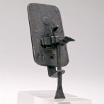
Early microscopes like the one made by Antoni van Leewenhoek, on display in Revealing the Invisible: The History of Glass and the Microscope, were used to examine the quality of cloth. Today microscopes are used in three important aspects of glass conservation: examination, research, and treatment.

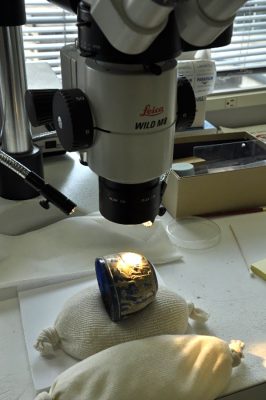
Conservator Astrid van Giffen uses a microscope to examine glass.
Examination
An important part of conservation is the extensive and detailed examination that each object undergoes before it is treated. This is done to determine the condition of the object as well as to discover details about how it was made. A stereomicroscope, which gives a 3-D image of an object, is just one of many tools used to help in examination of the small, fine details of glass.
Here are some examples of what details a closer look at an object can reveal:
-
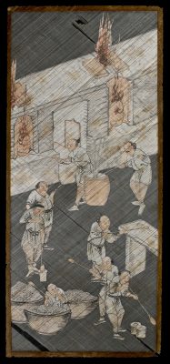
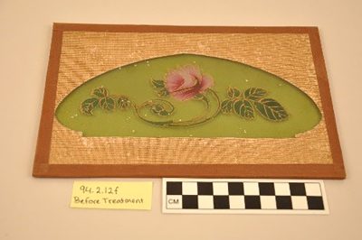
- Screen panel, China,1800-1899. Transfer from The Rakow Library. 2010.6.26.
-
- Screen panel detail under the microscope
This Chinese glass rod panel dated to the 19th century (above left) is made up of two panes of assembled glass rods with painted paper cutouts sandwiched in between. Under the microscope (above right), it became clear that most of the rods are hollow or partially hollow, which suggests that they were drawn or pulled. Read more about the conservation of this panel.
-
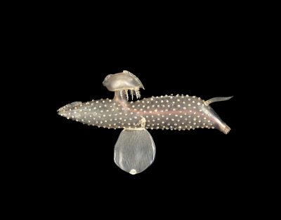
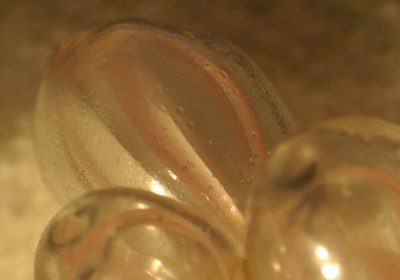
- Carinaria mediterranea (1885); Carinaria lamarckii (2016); Blashka Nr. 546, Leopold and Rudolf Blaschka, Dresden, Germany, 1885. Lent by Cornell University, Department of Ecology and Evolutionary Biology. L.17.3.63-102.
-
- Detail of Blaschka Nr. 546 under the microscope
At first glance it might look like all of the dots on Blaschka Nr. 546 (above left), on display in the current exhibition Fragile Legacy: The Marine Invertebrate Glass Models of Leopold and Rudolf Blaschka, could be fused to the surface, but under the microscope you can clearly see the glue (now yellowed and cracking) used to hold each one in place. Also apparent is the matte coating of the surface; this is especially noticeable where one of the dots has come off and we can see the uncoated glass underneath.
-
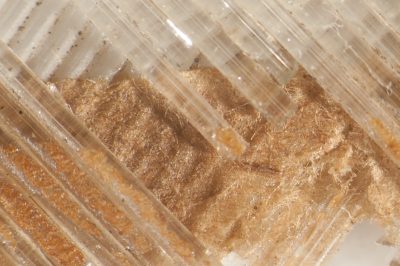
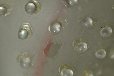
- Lampshade sample, Smith Brothers Decorating Co., New Bedford, Mass., 1890-1910. Gift of The New Bedford Glass Society. 92.4.12.
-
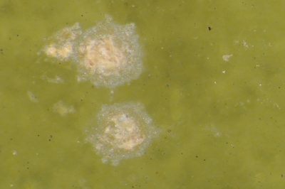
- Detail of lampshade sample under a microscope
This lampshade sample (above left) has some strange white spots on its surface. Under the microscope you could see that they are pits of glass deterioration (above right).
-
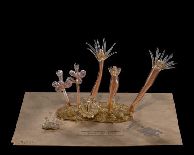
- Podocoryne carnea (1885); Podocoryna carnea (2016); Blaschka Nr. 173, Leopold and Rudolf Blaschka, Dresden, Germany, 1885. Lent by Cornell University, Department of Ecology and Evolutionary Biology. L.17.3.63-267.
-
- Detail of Blaschka Nr. 173 under a microscope
Early symptoms of atmospheric glass deterioration can be more easily seen under the microscope too, like the droplets on the inside of these glass polyps from Blaschka Nr. 173, also on display in Fragile Legacy.
-
- Examining the Morgan Cup (52.1.93) under a microscope
-
- Detail of the Morgan Cup (52.1.93) as seen under a microscope
Much of the surface of this Roman cameo glass, the Morgan cup on display in 35 Centuries of Glass, is heavily weathered, a type of glass deterioration that occurs when glass is buried. When glass deteriorates it starts from the surface and moves inward, unlike metal corrosion, which grows on top of the surface. This means that the surface of the weathering layer represents the original smooth surface of the glass; when this layer is lost, surface details are lost as well. In the image above, the white pitted surface on the left is where the original surface has been lost.
Research
More powerful microscopes, such as a scanning electron microscope (SEM), are sometimes used in research in collaboration with conservation scientists. An SEM scans a focused electron beam over a surface to create an image. Because this process is done in a vacuum chamber, only small, stable objects or samples can be examined. SEM is often coupled with energy-dispersive X-ray spectroscopy (EDS, EDX, or XEDS) which allows analysis of the elements in the glass and visually represents different compositions. This important analytical component usually requires the use of samples that are embedded in a resin, polished, and coated with a thin layer of carbon or gold, which is not always possible.
-
- Glass sample viewed through an SEM.
-
- Glass sample embedded in resin
-
- Glass sample before it is embedded in resin
This series of images shows a sample before it was embedded in resin (bottom right); the embedded and polished sample viewed with a reflected light microscope (top right); and viewed with SEM (above left). The reflected light microscope and SEM images show mixing of glasses with different compositions.

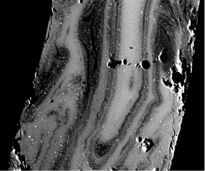
This is an SEM image (above) of the cross-section of a glass bead with an opaque white core and a pink translucent outer layer. A closer look at the rim of the cross-section reveals a thin layer of a slightly different composition indicating glass deterioration. This deterioration was not visible without the SEM.
Treatment
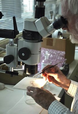
A stereomicroscope is extremely valuable for many different types of treatment. The magnification provided by working under the microscope allows more control over the process, allows better precision, and helps minimize the risk of unintentional damage.
When an object is broken into many pieces (below left), the correct placement of each fragment is crucial. Because many of the objects treated have been repaired in the past, an important first step in treatments is often cleaning off the old glues and fill materials. Cleaning the break edges is usually done under the microscope because even a tiny bit of extra glue can cause the fragment to be misaligned, which in turn will cause other fragments to be placed incorrectly.
-
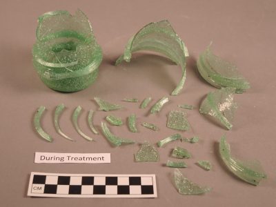
- Broken object prior to treatment
-
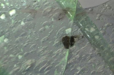
- Aligning the fragments under the microscope
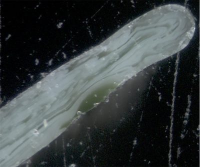
The alignment of the fragments is often checked under the microscope (above right); lining up inclusions, tiny bubbles, or streaks in the glass that cross the breaks helps ensure that each fragment is properly placed.
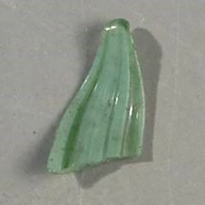
-
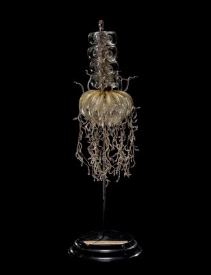
- Physophora magnifica (1885); Physophora hydrostatica (2016), Blaschka Nr. 213,Leopold and Rudolf Blaschka,Dresden, Germany, 1885. Lent by Cornell University, Department of Ecology and Evolutionary Biology. L.17.3.63-516.
-
- Reconstructing Nr. 213 under a microscope
-
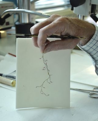
- A reconstructed tentacle of Nr. 213
The tentacles of Blaschka Nr. 213, on display in Fragile Legacy, were broken into many parts and were re-constructed under the microscope.
-
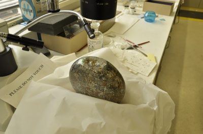
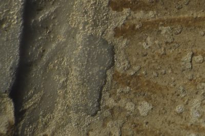
- A bottle (66.1.3) is prepared for cleaning.
-
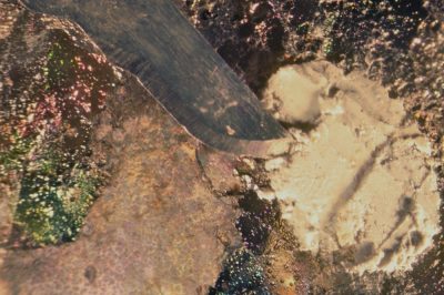
- A scalpel is used to remove putty from the bottle.
Cleaning objects with sensitive or fragile surfaces, such as this heavily weathered glass bottle, is also done under the microscope. In the photomicrograph (photograph taken through a microscope) above, a scalpel is being used to carefully remove a white putty that was stuck to and in the surface.

Revealing the Invisible: The History of Glass and the Microscope is open 9 am – 5 pm every day through March 19, 2017. This exhibition tells the stories of scientists’ and artists’ exploration of the microscopic world between the 1600s and the late 1800s. Unleash your sense of discovery as you explore the invisible through historic microscopes, rare books, and period illustrations, and take a #cellfie in the Be Microscopic interactive.